Introduction to cell organelles
Cell organelles is a core JC1 topic in H2 BIO A levels. This topic is one of the most important foundation to the entire H2 BIO syllabus. It explores the microscopic appearance of cells, it’s contents, their corresponding functions as well as their assigned names (plural/singular).
Materials for cell organelles

- 👆🏻 Quick video tips about the topic.
- Website showing the differences between prokaryote and eukaryote ribosomal proteins.
- Download diagrams (high resolution) – FREE!
- BUY notes ⌲ includes phrasing error corrections & review question solutions.
- Purchase notes (including phrasing error corrections & review question solutions) for cell organelles.
Phrasing errors
- The cell membrane is a …
- Nucleolus consists of DNA.
- Lysosomes contain lysozymes.
- The nuclear envelop is a bilayer.
- Mitochondria are sites of respiration and they produce energy.
Exam tips
- Centrioles are not found in most plants.
- Transport vesicles are different from secretory vesicles.
- Cisterna and crista refers to structures in different organelles.
- When asked to label, check arrow # and give appropriate singular or plural forms.
- It is not important to contrast animal and plant cells anymore but one should understand and be able to describe what structures makes plants unique e.g. chloroplasts and cellulose.
- Glycosylation of proteins begin in the RER vs. lipids in GA. What happens to the glycoproteins in GA is further/final chemical modifications as well as packing and sorting.
- Finally, many students do not spend enough time contemplating the concept of cell theory. Make sure you do as the idea of viruses being non-living is built on this very concept.
Calibration of microscopes to measure cell organelles Part 1
- Idea ⌲ You need to measure a structure you are looking at in the microscope to determine the actual length. In order to do so, someone thought why not use a ruler? However, if you use a ruler together with the sample, this presents with a logistical problem. Since every time the sample have to be prepared together with a ruler. After that it has to be washed. As such, a solution is to stick it to the ocular eyepiece instead. That way, you will constantly have a ruler that you can reference and use to measure the size of the structure you are seeing in the microscope. However…
- Problem ⌲ When you stick an ocular ruler into the eye piece, it remains static even though you increase magnification because you want to get a clearer view of the cellular details. As such, if the magnification changes, the unit length represented by the division of the ocular ruler will therefor have to change as well.
- Solution ⌲ Consequently, you need to calibrate what each division of your ocular ruler represents in length under each magnification. In order to do that, you use a stage micrometer that has a known length per division. That way, when you have a certain magnification, you can work out the actual length of a division on the ocular ruler by multiplying the number of divisions the structure occupies with the unit length at a particular magnification.
Calibration of microscopes to measure cell organelles Part 2
- Steps ⌲ Decide on which magnification you want to use. Then try to align the stage micrometer with the ocular micrometer as shown below using the same magnification.
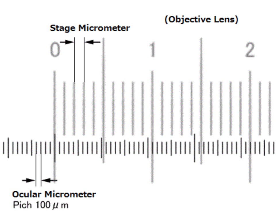
- Calculations ⌲ The stage micrometer total length will be = (unit length*) X (total # of divisions within the 2 meeting points). Once you get the total length (L), then your calculation of what is a unit length of the ocular ruler will be ⌲ (L) / (the total # divisions within the aligned 2 points). This will give you a unit length of the ocular ruler at the particular magnification that you used. * will be made known to you.